MAGNETIC MODULATION BIOSENSING (MMB) SYSTEM
MAIN GOAL
 Detection of biomarkers in low concentrations.
MOTIVATION
In recent years, biomarkers, such as proteins or nucleic acids, have become a very important tool for detection of various diseases. Even minor changes in biomarkers concentrations may be an indication of first signs of a disease. Usually, biomarkers are present at relatively low concentrations. Therefore, in order to detect such low concentrations, highly sensitive medical instruments are required.
CHALLENGES
1. High-sensitivity assays for biomarkers measurements have been developed, but they are usually done in laboratories, using complex and expensive equipment. From the other hand, to enable health care providers to make more timely decisions, biomarkers measurement need be performed at the patient’s bedside, but the devices used in these settings are associated with less precise and less sensitive performance.
 2. In many bioligical assays, the target protein is marked with a fluorescent marker. To see the fluorescent marker, one need to shine laser light on the solution. However, at low concentrations, only two or three molecules are positioned inside the detection volume and therefore the fluorescence signal is very weak which limits the minimum cocentration of detection.
MMB
In order to try and overcome the challenges , a novel high-sensitivity biomarker detection platform, named Magnetic Modulation Biosensing (MMB), has been developed in 2008 by Dr. Amos Danielli.
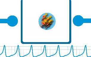
The MMB uniquely combines magnetic and fluorescent labeling to measure ultra-low biomarker concentrations: the target molecules are attached to magnetic beads. When magnetic forces are been activated, the beads concentrate from the entire solution into the detection volume. Instead of two-three molecules, there are thousands of molecules in detection area and the signal increases by orders of magnitude.
To eliminate the background noise , a modulation in magnetic field is generated by two electromagnets. As a result, the magnetic beads move from side to side, in and out of the laser beam spot. Every time they pass through the laser beam, they generate light.
DETECTION PROCESS
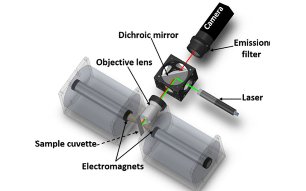
A laser beam is reshaped using two plano-convex lenses, reflected by a dichroic mirror, and focused by an objective lens onto the sample. The sample is a magnetic bead aggregate inside a rectangular glass cell positioned between two electromagnets that cause the bead aggregates to move. The laser excites the fluorophores in the sample, and the emitted fluorescence is collected by the same objective lens, filtered by an emission filter, and detected by a CCD camera or by a photomultiplier tube (PMT). A precision pinhole blocks the scattered photons from reaching the PMT.
SYSTEM CAPABILITIES
System capabilities have been demonstrated using a commercially-available assay for detection and quantification of human Interleukin-8 over several orders of magnitude. The results show that MMB has a 6-log dynamic range available for detection and is capable of detecting 0.08 ng/L of IL-8 in blood plasma, a sensitivity comparable to current state of the art laboratory assays.
The IL-8 experiments results were published in a paper at Sensors and Actuators B: Chemical. The paper, entitled: "High sensitivity detection of a protein biomarker interleukin-8 utilizing a magnetic modulation biosensing system" can be found in the Publications section, and at DOI: 10.1016/j.snb.2016.10.089.